Eps 1: Sutural Bone Is Bound To Make An Impact In Your Business
| Host image: | StyleGAN neural net |
|---|---|
| Content creation: | GPT-3.5, |
Host

Lily Woods
Podcast Content
The IF, when present, lies within the interfrontal suture , usually located anteriorly close to the nasal bones. The bone of the IF, being a secondary bone feature, has prompted interests which are likely wider in relevance with regard to the biology of metopic suture. A better understanding of IF bone formation could result in an increased knowledge about other bone formations within the cranial cavity, such as the Wormyian bones, and aspects of the biology of the skull suture.
Thus, better understanding the genetic and environmental factors influencing the IF bone formation could have implications for understanding the wormian bone formation. This study seeks a better understanding of the interactions between external mechanical forces and the bone deposition within sutures. Using a theoretical approach, we demonstrate that external mechanical forces are sufficient for the function of sutures in ways that replicate many in vivo features of bone deposition within the suture.
The fact that the deposition of bone occurs at the edges of the suture is influenced by external mechanical stimuli has been demonstrated on experimental systems . Apart from a genetic plan by default, the sutural growth is probably a function of specific parameters of the mechanical stimuli.2 The exact characteristics of the anabolic mechanical stimuli of the sutural bone growth are unknown. Genetic factors only exert nonspecific morphogenetic effects on intramembranous bone formation, and they determine only the outer limits and the increase in number of periods of growth.
Intramembranous bone formation is found during the skull growth, also found in the skull and the mandible, although composed of endochondral elements, and endochondral and intramembranous growth processes are occurring within the same bone. Endochondral bone formation may occur in bones associated with joint movements and in certain parts of the craniobase.
Craniofacial bones are connected with sutures that function as joints, absorbing impact forces and function as sites for intramembranous bone growth . Sutures are soft connective tissues that form joints between the bones of the skull. Cranial and facial sutures are connective tissue joints between the bones of the skull.
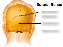
Wormian bones, also known as subsutural bones or cranial bones, are additional pieces of bone that may arise inside the suture of the skull. Wormian bones are small, irregular bones present inside a suture or femoris. Wormian bones are sometimes seen also inside the spinal sutures as well as coronal ones.
Placoderms constitute an older group in which dermal bones and sutures may relate to tissues observed in more descended groups, such as mammals . The appearance of Wormian bones is thought to result from disturbed osteogenesis/ossification, or a response to mechanical forces that impact the suture . In many cases, mechanical stresses resulting from muscle contraction produce large bending moments and consequently large bone strains adjacent to several cranial and facial sutures such as the zygomaticotemporal19,20,22,24 and reaction forces from the temporomandibular joint.23,44 In hindsight, the beam hypothesis was a much-needed step forward from the notion that craniofacial bones are incapable of stress-bearing and was proposed before the availability of experimental data on bone strain, primarily collected in the past two decades.
The facial deformations suggest that the sutures are capable of resisting mechanical stresses without excessively loading facial bones. This type of evidence is typically obtained by applying strain gauges, an engineering-based gauge used in structural mechanics, on the cortical surfaces of cranial bones, either adjacent to the sutures or directly above them, in animal models under mastication. Synchrotron microtomography confirmed the presence of the Sharpey fibers in peri-sutural bones.
The biomechanical model is, to our knowledge, the first attempt at building a theoretical link between external mechanical conditions and the cell events that lead to formation of the sutural bone. A biomechanical model is, to our knowledge, the first attempt to build a theoretical relationship between external mechanical conditions and cellular events resulting in sutural bone formation. There are two types of ossification, called intramembranous ossification, in which the marrow cells are differentiated into osteoblasts in an ossification centre immediately, with no previous cartilage formation, and endochondral ossification, in which the bone tissues are deposited by cartilage formation. The second is endochondral ossification, where bone tissue substitutes for the existing hyaline cartilage, such as in the formation of skull bases.
Bone tissue forms bones in the skull and jaw; particularly this occurs during only the developmental period, and also in the process of fracture repair. Compact bone forms the solid outer layer of all bones, surrounding a medullary cavity, or bone marrow. Bone tissues found in the periosteum, endosteum, sutures, and the periodontal membrane are examples of intramembranous bone formation .
Craniosynostosis typically involves the premature joining of one single skull suture, but can involve more than one of the sutures of the infants skull . Craniosynostosis is a condition that occurs at birth, where one or more joints made of solid tissue are closed too early , before the brain has fully formed in the baby. Cranial Sutures and Fontanels Open pop-up dialog box Close Cranial Sutures and Fontanels Cranial sutures and fontanels The joints made of strong, fibrous tissue that connect your babys bones to each other.
Usually, infancy, joints made of stronger material stay pliable, allowing the babys skull to expand as his or her brain grows. Prematurely fused septal joints running from front to back of the skull Top of skull Pressure on the head growing longer and narrower. Premature fusion of either of the coronal sutures running from each ear to the top of the skull can result in flattened foreheads on affected sides and ridged ones on the unaffected sides.
The risk of increased pressure within the skull with simple craniosynostosis is low when surgically fixing the sutures and the shape of the head. The disruption of the closure of interfontal suture via Ambn-Msx2 axis results in the thinning and enlargement of IF bones in mice . The existence of this population could provide yet another indication in favor of the fact that sutures are an active site for bone deposition during postnatal growth.