Eps 1097: how to use ultrasound
— The too lazy to register an account podcast
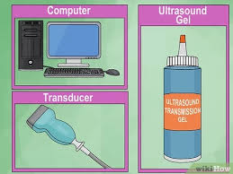
A computer uses the sound waves to create an image.
Ultrasound is not an ideal imaging technique for the air-filled bowel or organs obscured by the bowel.
Ultrasound has difficulty penetrating bone and can only see the outer surface of bony structures and not what lies within (except in infants who have more cartilage in their skeletons than older children or adults).
Host

Roger Marshall
Podcast Content
Ultrasound uses radio frequency sound waves to create an image of an object, such as a person's skin, blood vessels, organs or tissue. A licensed technologist will use a transducer or wand to send the sound wave. In this type of ultrasound examination, the rod that emits the sound waves and picks up the echoes is pressed against the skin surface and moved. While the sound waves reflect the organs and tissues, a computer translates the echo into the image.
In other cases, to get a good picture, the doctor must use a transducer placed in a tube connected to the neck or stomach, as in the case of a heart attack.
Ultrasound, also called ultrasound or sonography, is an imaging method that uses sound waves to produce an image of a body part. A computer program analyzes the echoes of the sound waves sent through the body and generates the image on a screen. Special ultrasound machines, known as Doppler flow machines, can show how fast blood flows towards vessels.
Ultrasound scans or sonographs use high frequency sound waves to look at the body. Unlike mammograms, which use radiation or X-rays, ultrasound exposes a region of the body that is of interest to him to high-frequency sound waves. Because ultrasound images are taken in real time, they can show how fast blood flows through blood vessels, such as the flow of blood from a blood vessel to an artery.
X-rays, the ionizing radiation exposure associated with ultrasound images, expose patients to ionized radiation. Ultrasound examinations subject the patient to high frequency sound waves and ionise the radiation as well as the radiation from the body surface.
Instead, ultrasound uses sound waves to generate images from inside the body and is effective in imaging the lungs and head because the sound is not well transported between the bones and the air in the body.
Ultrasound scans usually do not require special preparation and there are no known risks associated with ultrasound scans. It is a widely used clinical procedure because it is one of the few imaging methods in which a landmark at a certain level serves as a diagnostic reference. In cardiac ultrasound examinations, the boundary stone is typically defined as measuring the width of an intersection between the ventricles, such as the ring point and the mitral valve. Landmarks such as umbilical cord, cervix, uterus and ovaries are considered important for obstetric ultrasound.
In ultrasonic devices, users have to adjust the caliper to a desired location using a trackball, which makes the work even more complex. Moreover, the reliability of the measurement may suffer from subjective differences of opinion between users.
New ultrasound technology has also made it easier to obtain abdominal images if someone is obese or is difficult to scan for other reasons. In some cases, technicians can control the depth of the ultrasound signal so that the scan provides a better quality image for doctors to examine.
As ultrasound is painless and does not have harmful radiation, it is an excellent option for use in children. The relatively weak ultrasound waves can be used in a variety of ways, such as in the presence of blood, blood vessels and even clots.
If you have a painful kidney stone, firing strong ultrasound waves through your body can make the stone vibrate and break apart. Sometimes the powerful ultrasound wave is also used to destroy damaged parts of the body such as the heart, lungs, liver, kidneys and other organs.
Similarly, ultrasound waves can be used to clean machine parts that need to be cleaned in other ways, such as blood vessels, organs, and other body parts. Ultrasound is a form of body imaging in which sound waves are used to facilitate medical diagnoses.
Qualified ultrasound technicians are able to look into the body by means of ultrasound to answer questions that the attending physician can ask. Normally a radiologist will supervise the ultrasound test and report the results. Other types of doctors can also use ultrasound as a diagnostic tool: surgeons and emergency doctors use it at the bedside to assess abdominal pain and other ailments.
Doctors assess organs for infection, damage and disease, but more is discussed below, as is the use of ultrasound as a diagnostic tool.
Ultrasound is a safe, painless, non-invasive procedure that has been used in medicine since the 1950s. Unlike many other imaging techniques, ultrasound does not use radiation to produce an image of the inner body.
You may also hear the word 'sonography' used in connection with an ultrasound examination, but sonography refers to the ultrasound device used to create the sonogram. A sonographer is an experienced technician who operates the device, and you may have heard of it being used as a reference for ultrasound examinations. The technicians use a rod-shaped device, called a transducer, to send sound waves through body parts for examination.